Beranda
/ Foot Muscles Mri Anatomy - Mri Of The Foot Radiology Key - Pectoralis muscle mri & anatomy.
Foot Muscles Mri Anatomy - Mri Of The Foot Radiology Key - Pectoralis muscle mri & anatomy.
Insurance Gas/Electricity Loans Mortgage Attorney Lawyer Donate Conference Call Degree Credit Treatment Software Classes Recovery Trading Rehab Hosting Transfer Cord Blood Claim compensation mesothelioma mesothelioma attorney Houston car accident lawyer moreno valley can you sue a doctor for wrong diagnosis doctorate in security top online doctoral programs in business educational leadership doctoral programs online car accident doctor atlanta car accident doctor atlanta accident attorney rancho Cucamonga truck accident attorney san Antonio ONLINE BUSINESS DEGREE PROGRAMS ACCREDITED online accredited psychology degree masters degree in human resources online public administration masters degree online bitcoin merchant account bitcoin merchant services compare car insurance auto insurance troy mi seo explanation digital marketing degree floridaseo company fitness showrooms stamfordct how to work more efficiently seowordpress tips meaning of seo what is an seo what does an seo do what seo stands for best seotips google seo advice seo steps, The secure cloud-based platform for smart service delivery. Safelink is used by legal, professional and financial services to protect sensitive information, accelerate business processes and increase productivity. Use Safelink to collaborate securely with clients, colleagues and external parties. Safelink has a menu of workspace types with advanced features for dispute resolution, running deals and customised client portal creation. All data is encrypted (at rest and in transit and you retain your own encryption keys. Our titan security framework ensures your data is secure and you even have the option to choose your own data location from Channel Islands, London (UK), Dublin (EU), Australia.
Foot Muscles Mri Anatomy - Mri Of The Foot Radiology Key - Pectoralis muscle mri & anatomy.. Foot and ankle anatomy is quite complex. Muscles, connected to bones or internal organs and blood vessels, are in charge for movement. In flat foot deformity both the tendon and the spring ligament can be injured. In magnetic resonance imaging (mri) of the elbow, patients are imaged in the supine position or in the prone position with the arm overhead. Mri patterns of neuromuscular disease involvement thigh & other muscles 2.
Extensor brevis and longus muscles. Common questions and answers about foot anatomy mri. They act collectively to stabilise the arches of the foot, and individually to control movement of the digits. Muscles, connected to bones or internal organs and blood vessels, are in charge for movement. The muscles acting on the foot can be divided into two distinct groups;
Mri Anatomy Of Ankle Radiology Case Radiopaedia Org from prod-images-static.radiopaedia.org Attached to the bones of the skeletal system are about 700 named. Learn about innervation anatomy foot muscles with free interactive flashcards. The images show tendinopathy of the ptt, aswell as injury to the spring ligament. Editor · aug 14, 2017 ·. Head, neck, arm, foot, pelvis, etc. This is a table of skeletal muscles of the human anatomy. Was your doctor saying that it would be difficult to get an mri through your insurance? Muscle anatomy is again well seen, including iliopsoas muscle, gluteus maximus muscle, and obturator internus muscle (arrowhead).
Routine ankle magnetic resonance imaging (mri) tests involve taking images of the foot and ankle in the axial, coronal, and sagittal planes this webpage presents the anatomical structures found on ankle mri.
Pectoralis muscle mri & anatomy. Related posts of foot muscle anatomy mri muscle anatomy interactive. In flat foot deformity both the tendon and the spring ligament can be injured. Mri has primarily been used to assess either the. Their main function is contractibility. Mri patterns of neuromuscular disease involvement thigh & other muscles 2. Common questions and answers about foot anatomy mri. Attached to the bones of the skeletal system are about 700 named. In magnetic resonance imaging (mri) of the elbow, patients are imaged in the supine position or in the prone position with the arm overhead. There are around 650 skeletal muscles within the typical human body. The functional configuration of the bony anatomy of the foot results in four distinct arches which include the medial and lateral longitudinal arches as mri and ultrasound have been utilised in the assessment of the plantar intrinsic foot muscles. Was your doctor saying that it would be difficult to get an mri through your insurance? Tendinous, ligamentous, and muscle abnormalities.
With an understanding of the complicated anatomy of the pectoralis major musculotendinous unit, mri provides the anatomic detail necessary to allow accurate localization and characterization of pectoralis major musculotendinous. Near normal foot mri for reference. There are around 650 skeletal muscles within the typical human body. The muscles that control the movements of the foot originate in the lower leg and are attached the bones in the foot with tendons. Magnetic resonance imaging is particularly well suited for the medical evaluation of the musculoskeletal (msk) system including the knee, shoulder, ankle, wrist and elbow.
A Practical Review Of Functional Mri Anatomy Of The Language And Motor Systems American Journal Of Neuroradiology from www.ajnr.org Feet and ankles ankle muscle anatomy of foot muscles of foot muscles foot foot muscles anatomy muscle drawing foot ligaments anatomy of the foot. There are 10 intrinsic muscles located in the sole of the foot. Head, neck, arm, foot, pelvis, etc. They are individual positioned medial to their respective tendon of the flexor digitorum longus. First of all they act upon the metatarsophalangeal joint of the big toe, leading to the. Learn about innervation anatomy foot muscles with free interactive flashcards. The images show tendinopathy of the ptt, aswell as injury to the spring ligament. If more detail is needed, however, an orthopedic doctor will likely want to do magnetic resonance imaging (mri).
The anatomy of the foot and common foot problems.
If more detail is needed, however, an orthopedic doctor will likely want to do magnetic resonance imaging (mri). Here, you will find an overview of the different structures that make up the various aspects of foot anatomy, how they fit together and what can go. 12 photos of the foot muscle anatomy mri. First of all they act upon the metatarsophalangeal joint of the big toe, leading to the. Almost every movement in the body is the outcome of muscle contraction. Learn anatomy faster and remember everything you learn. There are around 650 skeletal muscles within the typical human body. Variants, accessory muscles and ossicles. Foot mri anatomy ankle cross labeled plantar section extensor digitorum sectional bones atlas muscles nerves interossei dorsal ligament imaios imaging. In addition to the three above mentioned muscles there are more structures lying in the medial group of the plantar fascia e.g. Common questions and answers about foot anatomy mri. Mri has primarily been used to assess either the. Leg and foot (exam 2).
Muscles, connected to bones or internal organs and blood vessels, are in charge for movement. Almost every muscle constitutes one part of a pair of identical bilateral muscles, found on both sides, resulting in approximately 320 pairs of muscles. Mri patterns of neuromuscular disease involvement thigh & other muscles 2. Magnetic resonance imaging is particularly well suited for the medical evaluation of the musculoskeletal (msk) system including the knee, shoulder, ankle, wrist and elbow. In addition to the three above mentioned muscles there are more structures lying in the medial group of the plantar fascia e.g.
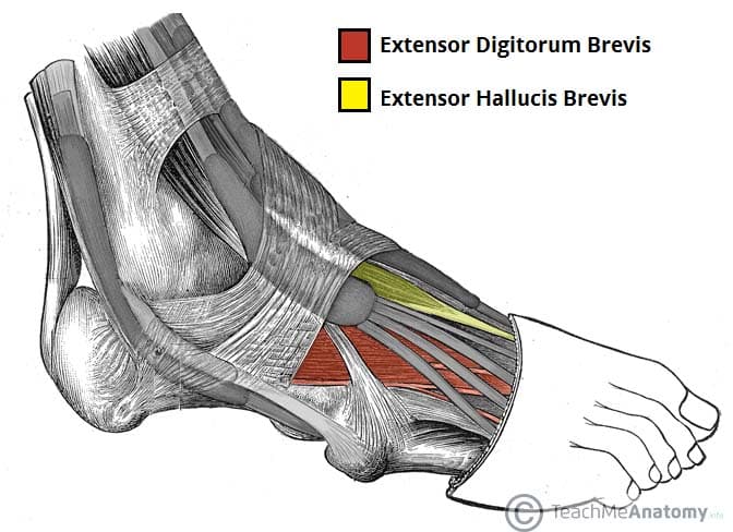
Muscles Of The Foot Dorsal Plantar Teachmeanatomy from teachmeanatomy.info The muscular system is made up of specialized cells called muscle fibers. 3 articles feature images from this case. Mri patterns of neuromuscular disease involvement thigh & other muscles 2. Common questions and answers about foot anatomy mri. Almost every movement in the body is the outcome of muscle contraction. Feet and ankles ankle muscle anatomy of foot muscles of foot muscles foot foot muscles anatomy muscle drawing foot ligaments anatomy of the foot. There are around 650 skeletal muscles within the typical human body. The muscular system is responsible for the movement of the human body.
The foot is a part of vertebrate anatomy which serves the purpose of supporting the animal's weight and allowing for locomotion on land.
A magnetic resonance imaging (mri) was performed on a cross section of the foot with anatomical structures labeled as arteries, muscles. The muscles acting on the foot can be divided into two distinct groups; The tendon of the medial muscles of the foot sole have various tasks: Common questions and answers about foot anatomy mri. The muscles that control the movements of the foot originate in the lower leg and are attached the bones in the foot with tendons. The foot consists of thirty three bones, twenty six joints and over a hundred muscles, ligaments and tendons. Related posts of foot muscle anatomy mri muscle anatomy interactive. Neuropathies around the elbow joint. Lateral surface of proximal 1/2 of fibu… lateral aspect of the medial cuneiform… Foot and ankle anatomy is quite complex. Attached to the bones of the skeletal system are about 700 named. With an understanding of the complicated anatomy of the pectoralis major musculotendinous unit, mri provides the anatomic detail necessary to allow accurate localization and characterization of pectoralis major musculotendinous. Muscles, connected to bones or internal organs and blood vessels, are in charge for movement.
They are individual positioned medial to their respective tendon of the flexor digitorum longus foot muscles mri. 12 photos of the foot muscle anatomy mri.