Beranda
/ Upper Leg Tendon Anatomy / Anatomy Of Leg Muscles And Tendons Anatomy Diagram Leg ... - Tendon, tissue that attaches a muscle to other body parts, usually bones.
Upper Leg Tendon Anatomy / Anatomy Of Leg Muscles And Tendons Anatomy Diagram Leg ... - Tendon, tissue that attaches a muscle to other body parts, usually bones.
Insurance Gas/Electricity Loans Mortgage Attorney Lawyer Donate Conference Call Degree Credit Treatment Software Classes Recovery Trading Rehab Hosting Transfer Cord Blood Claim compensation mesothelioma mesothelioma attorney Houston car accident lawyer moreno valley can you sue a doctor for wrong diagnosis doctorate in security top online doctoral programs in business educational leadership doctoral programs online car accident doctor atlanta car accident doctor atlanta accident attorney rancho Cucamonga truck accident attorney san Antonio ONLINE BUSINESS DEGREE PROGRAMS ACCREDITED online accredited psychology degree masters degree in human resources online public administration masters degree online bitcoin merchant account bitcoin merchant services compare car insurance auto insurance troy mi seo explanation digital marketing degree floridaseo company fitness showrooms stamfordct how to work more efficiently seowordpress tips meaning of seo what is an seo what does an seo do what seo stands for best seotips google seo advice seo steps, The secure cloud-based platform for smart service delivery. Safelink is used by legal, professional and financial services to protect sensitive information, accelerate business processes and increase productivity. Use Safelink to collaborate securely with clients, colleagues and external parties. Safelink has a menu of workspace types with advanced features for dispute resolution, running deals and customised client portal creation. All data is encrypted (at rest and in transit and you retain your own encryption keys. Our titan security framework ensures your data is secure and you even have the option to choose your own data location from Channel Islands, London (UK), Dublin (EU), Australia.
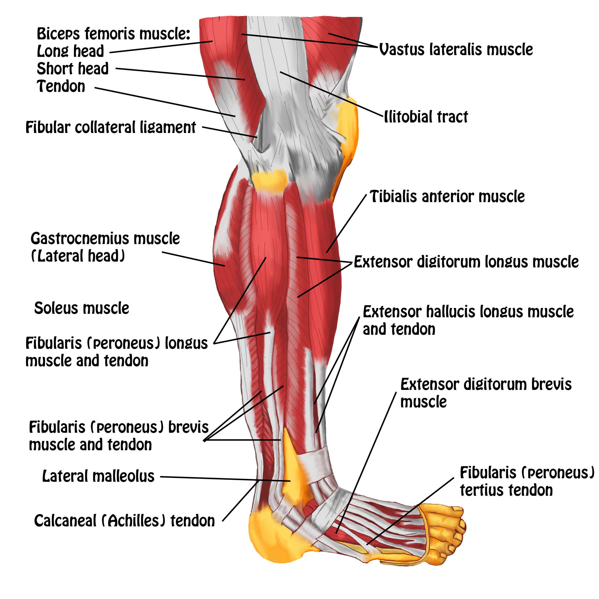
Upper Leg Tendon Anatomy / Anatomy Of Leg Muscles And Tendons Anatomy Diagram Leg ... - Tendon, tissue that attaches a muscle to other body parts, usually bones.. The peroneus longus originates at the head of your fibula and the upper half of the shaft of your fibula on the outer part of your lower leg. Tendons of the anterior compartment of the leg, the anterior tibial vessels, and the deep peroneal nerve pass under it. The lower leg is comprised of two bones, the tibia and the smaller fibula. Iliotibial band syndrome description the iliotibial band is the tendon attachment of hip muscles into the upper leg (tibia) just below the knee to the outer side of the front of the leg. We created an anatomical atlas of the upper limb, an interactive tool for studying the conventional anatomy of the shoulder, arm, forearm, wrist and hand based on an axial magnetic resonance of the entire upper limb.
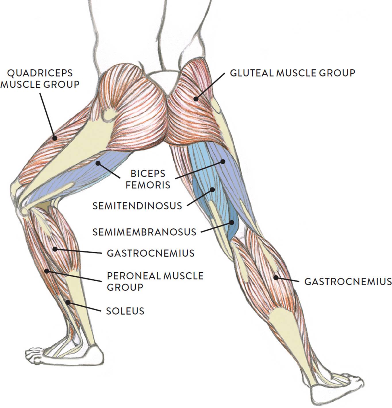
However, many reflex pathways are also active in the legs and foot. There is no real division between the core and the upper leg; Quadriceps tendon to base of patella and onto tibial tuberosity via the patellar ligament action: The large achilles tendon is the most important tendon for walking, running, and jumping. Do anatomy tracings over those to find the leg bones.
SMRT: Lower Leg & Foot - MASSAGE Magazine from www.massagemag.com Look for subcutaneous landmarks to figure out where the bones go. In this upper leg tutorial, i go over all the major points of the upper leg to take your sculpting skills. In human anatomy, the lower leg is that part of the lower limb that lies between the ankle and the knee. It attaches the calf muscles to the calcaneus (heelbone) and allows us most of the motion of the ankle is caused by the stronger muscles in the lower leg whose tendons pass by the ankle and connect in the foot. Do anatomy tracings over those to find the leg bones. We created an anatomical atlas of the upper limb, an interactive tool for studying the conventional anatomy of the shoulder, arm, forearm, wrist and hand based on an axial magnetic resonance of the entire upper limb. Information on the central tendon of the diaphragm by the anatomyzone daily feed. Tendons of the anterior compartment of the leg, the anterior tibial vessels, and the deep peroneal nerve pass under it.
Learn the origin/insertion, functions & exercises for the leg rotating your upper leg and pelvis to the inside or outside of your body's center line.
The nerve signals in these reflexes come from stretch receptors located in the joints, ligaments reflexes help to maintain proper muscle tone, balance, and responsiveness of the legs and feet to stimuli such as stepping on a sharp object. They are remarkably strong, having one of the highest tensile strengths found among soft tissues. Tendons are fibrous cords attached to muscles and bone. Try this movement out by standing on one foot with the other leg. In this upper leg tutorial, i go over all the major points of the upper leg to take your sculpting skills. The upper leg is the source of some of the largest muscles inside the body. The lower leg is comprised of two bones, the tibia and the smaller fibula. The pads of the machine are situated at the achilles tendon. Information on the central tendon of the diaphragm by the anatomyzone daily feed. Subscribe to learn interesting facts about the human body every day. To download this image, create an account. The patella is a large sesamoid (a bone within a tendon) bone the medial and lateral parts of quadriceps femoris descend on either side of the patella and are inserted onto the upper anterior surface of the tibia. There are several muscles which lie on the outside of your lower leg and are collectively known as the peroneal muscles (figure 1).
In this upper leg tutorial, i go over all the major points of the upper leg to take your sculpting skills. Tendinous sheath of right flexor pollicis longus radial bursa. Extends leg at knee in quad group. The appendicular skeleton includes the bones of the shoulder girdle, the upper limbs, the pelvic girdle, and the lower limbs. Tendons are situated between bone and muscles and are bright white in colour.
Musculature Stock Images, Royalty-Free Images & Vectors ... from thumb7.shutterstock.com Learn vocabulary, terms and more with flashcards, games and other study tools. Hands are outstretched, holding onto the handles of the bench. The nerve signals in these reflexes come from stretch receptors located in the joints, ligaments reflexes help to maintain proper muscle tone, balance, and responsiveness of the legs and feet to stimuli such as stepping on a sharp object. Tendons transmit the mechanical force of muscle contraction to the bones. Learn the origin/insertion, functions & exercises for the leg rotating your upper leg and pelvis to the inside or outside of your body's center line. The anatomical basis of clinical practice. Spicermanyt at checkout for 40% off this tutorial! Enters the anterior compartment of the leg through a gap in the upper part of the interosseous membrane.
Extends leg at knee in quad group.
It blends with the fibrous pericardium above, helping to. Information on the central tendon of the diaphragm by the anatomyzone daily feed. By spicer mcleroy in tutorials. The appendicular skeleton includes the bones of the shoulder girdle, the upper limbs, the pelvic girdle, and the lower limbs. It is the largest tendon of the parts of leg. However, many reflex pathways are also active in the legs and foot. There is no real division between the core and the upper leg; It attaches the calf muscles to the calcaneus (heelbone) and allows us most of the motion of the ankle is caused by the stronger muscles in the lower leg whose tendons pass by the ankle and connect in the foot. Enters the anterior compartment of the leg through a gap in the upper part of the interosseous membrane. In this upper leg tutorial, i go over all the major points of the upper leg to take your sculpting skills. Upper leg, knee, lower leg, ankle, and foot. The anatomical basis of clinical practice. Subscribe to learn interesting facts about the human body every day.
Subscribe to learn interesting facts about the human body every day. Try this movement out by standing on one foot with the other leg. The tendons that control movement in your hands, wrists and fingers run through your forearm. Lie prone on a hamstring curl machine. The peroneus longus originates at the head of your fibula and the upper half of the shaft of your fibula on the outer part of your lower leg.
Muscles of the Leg and Foot - Classic Human Anatomy in ... from doctorlib.info What is a peroneal tendon rupture? Synovial tendon sheaths of right fingers. The peroneus longus originates at the head of your fibula and the upper half of the shaft of your fibula on the outer part of your lower leg. However, many reflex pathways are also active in the legs and foot. And it is also critical to the walking process. The tendons that control movement in your hands, wrists and fingers run through your forearm. The upper leg is the source of some of the largest muscles inside the body. Collectively, the muscles in this area plantarflex and invert the foot.
Enters the anterior compartment of the leg through a gap in the upper part of the interosseous membrane.
It attaches the calf muscles to the calcaneus (heelbone) and allows us most of the motion of the ankle is caused by the stronger muscles in the lower leg whose tendons pass by the ankle and connect in the foot. To describe the mechanical properties of tendons. Enters the anterior compartment of the leg through a gap in the upper part of the interosseous membrane. Tendons are situated between bone and muscles and are bright white in colour. However, many reflex pathways are also active in the legs and foot. And it is also critical to the walking process. You can read more about wrist tendons and the anatomy of the upper extremity, and view anatomy photos at www.handcare.org. The leg is composed of five distinct sections: To download this image, create an account. The nerve signals in these reflexes come from stretch receptors located in the joints, ligaments reflexes help to maintain proper muscle tone, balance, and responsiveness of the legs and feet to stimuli such as stepping on a sharp object. The lower leg is comprised of two bones, the tibia and the smaller fibula. Look for subcutaneous landmarks to figure out where the bones go. The thigh and leg bones articulate at the knee joint that is protected and enhanced by the patella bone that supports the quadriceps tendon.